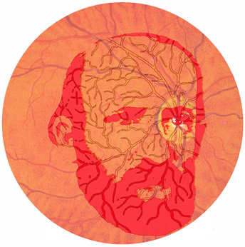
Heinrich Müller1820–1864
Helmholtz, who invented the ophthalmoscope in 1851, remarked: “The ground of the eye looks reddish, and indeed first of all round about the optic nerve of a somewhat clear, light-red, the darker, on the contrary, the farther you pass from that place”. In the same year, direct observation of the retinas of frogs by Heinrich Müller also led to descriptions of its reddish colour: “The rods of frogs appear somewhat reddish, where they lie in a sufficient density over one another, and one can see single rods alternatively colourless or coloured, depending upon whether they are lying or upright”. Müller was less interested in the colour of the retina than in its conformation. After medical training he moved to the University of Würzburg where he became professor of anatomy in 1858. He carried out most of his experimental studies there, but he travelled to Italy for health reasons in 1850 and 1851. It was there that he was led to the study of marine biology and he made observations on retinal histology in many species. Müller investigated the microanatomical structure of the retina in order to determine the location of the receptor elements. Achromatic microscopes were exposing unexpected detail in both the structure within the retina and the orientation of the likely receptors: they were pointing away from the incoming light rather than towards it, as had long been assumed. Müller’s experiments were not only microscopic. He was able to examine characteristics of the Purkinje tree (the visibility of shadows cast by retinal blood vessels) to establish their location in front of the receptor layer. He planned to write a book on the histology of the retina, but his early death denied him of this. Müller devoted his experimental life to studies of the retina and accomplished a remarkable amount in his short life; alas, he did not realise the potential that had been displayed. He is shown in a view of the retina through an ophthalmoscope and also a diagram of the Purkinje tree.
