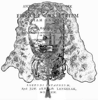
Antonius van Leeuwenhoek1632–1723
While the microscopes of Hooke and Leeuwenhoek bared the cell before the human eye, they did little to expose its fine structure. This was to await the development of achromatic microscopes in the early 19th century. Leeuwenhoek applied a simpler magnifier of greater power than Hooke’s; his descriptions of animal tissue were published in letters to the Royal Society. The collected letters were published as a book, the title page of which is shown, together with a portrait of Leeuwenhoek and his simple magnifier. Starting in 1674, Leeuwenhoek gave accounts of several animal cells, including nerve fibres. In the following year he wrote: “I represent to my self a tall Beer‑glass full of Water: This Glass I imagine to be one of the filaments of the Optic Nerve, and the Water in the Glass to be the globuls of which the filaments of that Nerve are made up, and then, the Water in the Glass being toucht on its surface with the finger, that to this contact did resemble the action of the visible object upon the Eye, whereby the outermost globuls of the fibres in the Optic Nerve next to the Eye are toucht. This contact of the Water made by the finger cannot be said to touch and move only the surface of the Water, but we must also grant, that all the water in the Glass is moved thereby, and that even the bottom of the Glass comes to suffer, and to be more pressed by it, than it was before the finger touched the Water, and that also all the parts of the Water are moved thereby. This motion then of the Water, said to be made by the contact of the finger, I imagine to be like the motion of a visible object made upon the soft Globuls, that lie at the end of the Optic Nerve next the Eye, which outermost globuls do communicate the like motion to the other globuls so as to convey it to the Brain.” Relatively little attention was paid to these observations because the resolving power of the microscopes was poor.. Microanatomy was not advanced greatly in the 18th century, and the use of the microscope was often disparaged. Despite these technical limitations, Leeuwenhoek provided the first microscopic observation of nerve fibres, about one century before the more accurate description published in 1781 by Felice Fontana (1730-1805).
