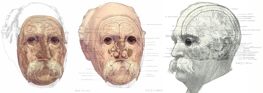
William Macewen1848–1924
Macewen was a general surgeon who pioneered operative methods for rickets, inguinal , bone defects, and otological problems, among others. Although much influenced Lister’s antiseptic procedures, he helped pioneer the alternative aseptic approach. operations were conducted in Glasgow, where he was to become Regius Professor of Surgery, a chair previously occupied by Lister. His brain surgery was based on the theories of Hughlings Jackson and Ferrier as well as the observations of Broca. It is usually claimed that Alexander Hughes Bennett (1848-1901) and Rickman John Godlee (1849-1925) carried out the first operations in 1884 to remove brain tumours based on localising signs, but that honour is rightly accorded Macewen. Prior to 1884 Macewen carried out seven such operations on the brain and eight on the spinal cord; in the former “cerebral localisation of function guided the operator to particular lesions”. He was accorded many honours for his pioneering work as a brain surgeon, and also for his work in otolaryngology, general surgery, and nurse training. Amongst these, he was knighted in 1902 and elected President of the British Medical Association in 1922. He was born on the Isle of Bute, where he is also buried. Macewen’s portrait is presented three times, in combination with images from his Atlas of Head Sections(1893). The Atlas is comprised of photographs and diagrams derived from 53 coronal, sagittal and horizontal sections of brains in frozen heads. The Plates showing the sections were presented as “an aid to cephalic topography” because the conformation of the brain in situ differs from that when removed from the head. Macewen is shown in Plate No. 1 with the photograph on the left and the diagram in the centre. The description is “CORONAL SECTION, viewed from before, passing through frontal lobes, posterior aspects of the eyeballs, the ethmoidal cells, antrum of Highmore, turbinated bones, buccal cavity and tongue.” He is also shown in profile combined with a diagram of a sagittal section of the brain: “SAGITTAL SECTION of the left side of the head… passing through outer half of orbit, malar bone, coronoid process, and inmost tip of condyle of lower jaw, styloid process and outer border of occipital condyle, showing eyeball with ocular muscles, temporal fossa, tympanic orifice of Eustachian tube, tensor tympani muscle, cochlea and facial nerve, upper portion of fissure of Rolando, operculum, island of Reil, portions of fissure of Sylvius, frontal, parietal, occipital and temporal lobes.”
