Marcello Malpighi1628–1694
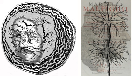
The application of early microscopes to biological specimens was a huge leap assisting understanding, but the microscopes themselves were neither powerful nor free from optical aberrations; the techniques for preparing specimens for observation were also wanting. The gross structures of the nervous system had been examined with the naked eye, but a new world was exposed by the microscope, and this world was accurately examined by Malpighi. This led him to the momentous discovery of blood capillaries thus putting Harvey’s hypothesis of blood circulation on a firm anatomical footing. He is shown on the left in his diagram of the developing chick in its egg. On the right his portrait is combined with the tracheal system of the silk worm and the title page of his medical and anatomical works. As a consequence of the many structures he examined microscopically, Malpighi is often regarded as the first histologist. In addition to chick embryos, where he provided one of the first accurate descriptions of development in the nervous system, he examined the skin (which now has a Malpighian layer), taste buds on the tongue, liver, kidneys (where he discovered Malpighian corpuscles). He received his medical education at the University of Bologna, and held posts in Pisa, Messina, Bologna and Rome. Malpighi applied to the interpretations of physiology the mechanistic concepts derived from the Galileian school; microscopic organization of living tissues was seen as based on the functioning of a multitude of minute machines. Like Galileo, he was strongly involved in defending the “new science” against the criticisms of the traditional milieu. Many of his observations were reported as letters to the Royal Society of London, to which he was elected as a Fellow.
